
CHRIS D. MELETIS, ND
Autoimmunity is a complex disease state with multiple factors contributing to its expression. Autoimmune disease is not simply a physical ailment. An emotional component may also be involved in the etiology of the condition. Our thoughts are capable of creating complex shifts in our biochemistry. In the case of autoimmune disease, thoughts can impact autoimmunity by contributing to dysfunctional immune system states. For example, someone who feels guilty or angry at themselves can experience autoimmune reactions consistent with “punishing” themselves for what they believe they did wrong. Paying attention to the power of our thoughts, addressing dysfunction at multiple levels of physiology (physical, emotional, and psychological), and subsequently quieting the immune system’s unnecessary reactivity could be the goal for many of those suffering from autoimmunity and allergies.
Because so many things can affect autoimmunity, I will limit the discussion to 4 key factors that will benefit the majority of autoimmune patients. These factors include stress, adrenal function, NAD+, and vitamin D.
A Brief Review
The following are some additional considerations to keep in mind when treating autoimmune conditions.
- Mitochondria – Mitochondrial dysfunction is involved in autoimmune diseases, including systemic lupus erythematosus (SLE),1 rheumatoid arthritis (RA),2 and Hashimoto’s thyroiditis.3 In autoimmunity, mitochondrial respiratory chain complexes may change, and the body is unable to eliminate damaged mitochondria.3,4 Coenzyme Q10, which supports mitochondrial health, has been shown to improve the health of patients with RA,5 antiphospholipid syndrome,6 psoriasis (together with vitamin E and selenium),7 and diabetes.8
- Nitric oxide homeostasis – Nitric oxide (NO) is a chemical produced in the body that’s important to many biological processes. Rodent models have shown that NO bioavailability is reduced in autoimmune diseases like RA.9 Impaired NO metabolism has also been associated with the cardiovascular risk and vascular inflammation that occurs in RA.10,11
- Viruses– Viral infections may trigger the development of autoimmune diseases. For example, encephalomyocarditis infection can trigger autoimmune diabetes.12 Epstein-Barr virus (EBV) can trigger Graves’ disease, multiple sclerosis (MS), and SLE.13
- Hormones – Hormonal changes are involved in the developing and worsening of autoimmunity, especially in women during perimenopause and in their postmenopausal years. Alterations in hormones during menopause can influence inflammatory processes that increases susceptibility to immune system dysfunction. While women’s neutrophil percentages decline after the age of 50, their lymphocyte percentages concurrently increase. This combination leads to an increased risk of lymphocyte-mediated autoimmune disease during perimenopause.14 Correcting the balance of hormones such as estrogen, progesterone, and dehydroepiandrosterone (DHEA) can be beneficial for women experiencing autoimmunity. Research indicates that supplementation with DHEA in autoimmune disorders like SLE can also be beneficial.15
- The endocannabinoid system – The endocannabinoid system may regulate autoimmunity as evidenced by promising animal studies using cannabidiol (CBD). In rodent studies, CBD reduced autoimmune hepatitis,16 suppressed the development of type-1 autoimmune diabetes,17 and resulted in significant improvement in autoimmune myocarditis.18
- Food allergies and intolerances– Impaired intestinal permeability, also known as “leaky gut,” can lead to a vicious cycle in IgE- and non-IgE mediated food reactions. Leaky gut can be one of the many factors contributing to autoimmune disorders.19,20 Testing food allergies, intolerances, and implementing an elimination diet (if necessary) can be an important part of healing for a patient diagnosed with autoimmunity.
Stress
It is estimated that up to 80% of autoimmune patients experienced excessive emotional stress before the onset of their disease.21 Stress both precedes the development of autoimmune diseases and worsens symptoms.22 It becomes a vicious cycle because the onset of autoimmune disease also tends to cause a patient to experience additional stress, which in turn exacerbates and perpetuates the disease. Research is ongoing; however, there are many relevant studies that support the connection between stress and autoimmunity. For example:
- Epidemiological and animal models suggest that psychological stressors are involved in the development of type-1 diabetes mellitus.23 Early life stress during the ages of 5 to 9 is a strong risk factor for the development of type-1 diabetes.24
- Post-traumatic stress disorder and/or trauma both increase the incidence of SLE. Both conditions also worsen physical symptoms of the disease.25,26
- Patients with MS have reported experiencing unusually high amounts of stress up to 1 year before their diagnosis.27 In a study that compared MS and non-MS patients, the incidence of reported high stress levels was almost 42% greater in the MS patients.27 This correlation was especially pronounced in the 6 months leading up to diagnosis.27
Researchers have proposed that the relationship between stress and autoimmune diseases could be due to mechanisms of action that include the hypothalamic-pituitary-adrenal axis and the nervous system’s effects on immune cells.23 Chronic stress can also cause dysbiosis of the intestinal microbiota, which has been shown to cause leaky gut and contribute to the development of autoimmunity.22
Psychological stress can also reactivate viruses linked to autoimmune diseases. For example, stress is a known trigger for EBV, and EBV reactivation during times of stress further weakens cellular immunity.28 Some individuals are more susceptible to EBV reactivation, and this virus has been linked to multiple autoimmune diseases, autoimmune-like conditions, and chronic fatigue syndrome/myalgic encephalitis.28
Adrenal Function
Psychological stress can also wreak havoc on the adrenal glands. Adrenal gland dysfunction can manifest as either Addison’s disease, Cushing’s syndrome, or what functional medicine providers typically call “adrenal fatigue.” Addison’s disease is an autoimmune condition that is caused when immune cells target the adrenal glands and results in low cortisol levels.29,30 Cushing’s syndrome is marked by high cortisol levels due to the overproduction of cortisol or from taking corticosteroid medications.
Adrenal fatigue is more common than Addison’s or Cushing’s diseases. In adrenal fatigue, cortisol levels become dysregulated. This can present as being low in the morning and high in the evening. Adrenal fatigue is a subclinical form of adrenal insufficiency.
One way to treat autoimmune patients with adrenal fatigue is with low-dose hydrocortisone. On a non-stressful day, healthy adrenal glands produce roughly 20 mg of cortisol. A good dosage of hydrocortisone for patients under stress is 5 mg per day. The timing of the dose will depend on salivary hormone testing. If the patient’s morning cortisol level is low, that will be the best time for them to take the medication. If cortisol levels don’t drop until the afternoon, dosing can be delayed until then but should not be taken too close to bedtime. You can combine hydrocortisone with adaptogens like Rhodiola rosea and Withania somnifera for increased benefit.
In addition to hydrocortisone, CBD can help support autoimmune patients that also are experiencing dysregulated adrenal function. CBD directly affects autoimmune markers, can calm stress, and can ease agitation.31,32 Stress-relief practices, like mindfulness meditation and mindfulness-based stress reduction (MBSR) techniques, can also be helpful. Mindfulness-based practices have shown benefits for autoimmune patients as evidenced by lowered urinary cortisol levels, reduced anxiety, and improved biological and immunological markers related to autoimmunity.33
ooted in core
Buddhist principles that all physiological suffering arises from a judgmental mind
NAD+
Nicotinamide adenine dinucleotide (NAD+) is a metabolic intermediate used as a cofactor by many important enzymes and is needed for over 500 biochemical pathways within the body. NAD+ is used for ATP production, the citric acid cycle, and metabolism. It also regulates inflammation and immune system health.
NAD+ protects against the inflammation that occurs during autoimmune conditions as well as inflammation that can occur due to aging.34 For example, in animal studies, NAD+ suppresses the development of experimental autoimmune encephalomyelitis (EAE), a model of multiple sclerosis.35 Several rodent experiments using EAE models have found that raising NAD+ levels led to significant improvements, including decreased expression of inflammatory cells and lower levels of pro-inflammatory cytokines, demyelination, nerve injuries, and motor function abnormalities.36–38 Increased NAD+ also led to a slight delay in the onset of the disease.36–38 One mechanism researchers have highlighted is that NAD+ likely affects autoimmunity through its role in regulating the differentiation of CD4+ T cells and by inducing the production of the anti-inflammatory cytokine IL-10 by T helper type 1 (Th1) cells.35
Raising patients’ NAD+ levels may also reduce autoimmunity due to NAD+’s effects on the glycoprotein CD38.34 A complex molecule that can act as an NAD-depleting enzyme or receptor, CD38 has a number of roles in immune system function, including regulating cell differentiation and modulating inflammation by influencing cytokine release.34 Declining NAD+ levels during aging correlate with an increased expression and activity of CD38.39
Increasing levels of NAD+ can be accomplished with nicotinamide riboside (NR) supplementation, which is safe even at higher doses.40 In one study, monocytes examined from patients with SLE who were supplemented with NR had a reduced type 1 interferon response compared with controls.41 Type 1 interferons play an important role in the pathogenesis of autoimmunity, and the change in patient response was attributed to raising patient NAD+ concentrations.42 There is also an indication from animal and human studies that supplementation with NAD+ precursors may be beneficial in RA, type 1 diabetes, and other autoimmune diseases.40
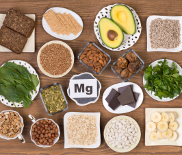
Vitamin D
Adequate vitamin D levels play an important role in maintaining healthy immune responses. The vitamin D receptor has been found on many immune cells, including monocytes, dendritic cells, and activated T cells.46 This indicates that it plays a regulatory role in preventing autoimmunity.46 Cell culture studies further indicate vitamin D blocks pro-inflammatory activity from CD4+ T cells by influencing their production of pro-inflammatory cytokines IL-2, interferon (IFN)-γ, and tumor necrosis factor-α.47,48
Studies in human patients with MS, type 1 diabetes, and SLE have found that patients with autoimmune diseases have lower serum concentrations of vitamin D compared to healthy controls.46 Epidemiological evidence indicates that the incidence of autoimmune diseases is also higher for people who live in regions with less ultraviolet light exposure from the sun.46 In some cases, lower vitamin D levels were also associated with MS relapses and may be involved in the progression and severity of the disease.49
Key Takeaways
The body seeks harmony and balance. It wants to not only exist but to thrive. Sometimes, we must push the body towards the path to health and healing. In autoimmune disease, this involves reducing stress, nourishing the adrenals, supporting NAD+ production, and ensuring adequate vitamin D.
[Refs]- Rai P, Janardhan KS, Meacham J, et al. IRGM1 links mitochondrial quality control to autoimmunity. Nat Immunol. 2021;22(3):312-321.
- Jaiswal KS, Khanna S, Ghosh A, et al. Differential mitochondrial genome in patients with Rheumatoid Arthritis. Autoimmunity. 2021;54(1):1-12.
- Zimmermann FA, Neureiter D, Feichtinger RG, et al. Deficiency of respiratory chain complex I in Hashimoto thyroiditis. Mitochondrion. 2016;26:1-6.
- Da Sylva TR, Connor A, Mburu Y, et al. Somatic mutations in the mitochondria of rheumatoid arthritis synoviocytes. Arthritis Res Ther. 2005;7(4):R844-851.
- Nachvak SM, Alipour B, Mahdavi AM, et al. Effects of coenzyme Q10 supplementation on matrix metalloproteinases and DAS-28 in patients with rheumatoid arthritis: a randomized, double-blind, placebo-controlled clinical trial. Clin Rheumatol. 2019;38(12):3367-3374.
- Pérez-Sánchez C, Aguirre M, Ruiz-Limón P, et al. Ubiquinol Effects on Antiphospholipid Syndrome Prothrombotic Profile: A Randomized, Placebo-Controlled Trial. Arterioscler Thromb Vasc Biol. 2017;37(10):1923-1932.
- Kharaeva Z, Gostova E, De Luca C, et al. Clinical and biochemical effects of coenzyme Q(10), vitamin E, and selenium supplementation to psoriasis patients. Nutrition. 2009;25(3):295-302.
- Brauner H, Lüthje P, Grünler J, et al. Markers of innate immune activity in patients with type 1 and type 2 diabetes mellitus and the effect of the anti-oxidant coenzyme Q10 on inflammatory activity. Clin Exp Immunol. 2014;177(2):478-482.
- Palma Zochio Tozzato G, Taipeiro EF, Spadella MA, et al. Collagen-induced arthritis increases inducible nitric oxide synthase not only in aorta but also in the cardiac and renal microcirculation of mice. Clin Exp Immunol. 2016;183(3):341-349.
- Mangoni AA, Tommasi S, Sotgia S, et al. Asymmetric dimethylarginine: a key player in the pathophysiology of endothelial dysfunction, vascular inflammation and atherosclerosis in rheumatoid arthritis? Curr Pharm Des. 2021;27(18):2131-2140.
- Crilly MA, McNeill G. Arterial dysfunction in patients with rheumatoid arthritis and the consumption of daily fruits and daily vegetables. Eur J Clin Nutr. 2012;66(3):345-352.
- Stafford JD, Yeo CT, Corbett JA. Inhibition of oxidative metabolism by nitric oxide restricts EMCV replication selectively in pancreatic beta-cells. J Biol Chem. 2020;295(52):18189-18198.
- Nagata K, Hayashi K. Epstein-Barr Virus Reactivation-Induced Immunoglobulin Production: Significance on Autoimmunity. Microorganisms. 2020;8(12):1875.
- Chen Y, Zhang Y, Zhao G, et al. Difference in Leukocyte Composition between Women before and after Menopausal Age, and Distinct Sexual Dimorphism. PLoS One. 2016;11(9):e0162953.
- Sahu P, Gidwani B, Dhongade HJ. Pharmacological activities of dehydroepiandrosterone: A review. Steroids. 2020;153:108507.
- Hegde VL, Nagarkatti PS, Nagarkatti M. Role of myeloid-derived suppressor cells in amelioration of experimental autoimmune hepatitis following activation of TRPV1 receptors by cannabidiol. PLoS One. 2011;6(4):e18281.
- Weiss L, Zeira M, Reich S, et al. Cannabidiol arrests onset of autoimmune diabetes in NOD mice. Neuropharmacology. 2008;54(1):244-249.
- Lee WS, Erdelyi K, Matyas C, et al. Cannabidiol Limits T Cell-Mediated Chronic Autoimmune Myocarditis: Implications to Autoimmune Disorders and Organ Transplantation. Mol Med. 2016;22:136-146.
- Teshima CW, Meddings JB. The measurement and clinical significance of intestinal permeability. Curr Gastroenterol Rep. 2008;10(5):443-449.
- Arrieta MC, Bistritz L, Meddings JB. Alterations in intestinal permeability. Gut. 2006;55(10):1512-1520.
- Stojanovich L, Marisavljevich D. Stress as a trigger of autoimmune disease. Autoimmun Rev. 2008;7(3):209-213.
- Ilchmann-Diounou H, Menard S. Psychological Stress, Intestinal Barrier Dysfunctions, and Autoimmune Disorders: An Overview. Front Immunol. 2020;11:1823.
- Sharif K, Watad A, Coplan L, et al. Psychological stress and type 1 diabetes mellitus: what is the link? Expert Rev Clin Immunol. 2018;14(12):1081-1088.
- Hägglöf B, Blom L, Dahlquist G, et al. The Swedish childhood diabetes study: indications of severe psychological stress as a risk factor for type 1 (insulin-dependent) diabetes mellitus in childhood. Diabetologia. 1991;34(8):579-583.
- Roberts AL, Malspeis S, Kubzansky LD, et al. Association of Trauma and Posttraumatic Stress Disorder With Incident Systemic Lupus Erythematosus in a Longitudinal Cohort of Women. Arthritis Rheumatol. 2017;69(11):2162-2169.
- Mills SD, Azizoddin D, Racaza GZ, et al. The psychometric properties of the Perceived Stress Scale-10 among patients with systemic lupus erythematosus. Lupus. 2017;26(11):1218-1223.
- Grant I, Brown GW, Harris T, et al. Severely threatening events and marked life difficulties preceding onset or exacerbation of multiple sclerosis. J Neurol Neurosurg Psychiatry. 1989;52(1):8-13.
- Kerr JR. Epstein-Barr virus (EBV) reactivation and therapeutic inhibitors. J Clin Pathol. 2019;72(10):651-658.
- Betterle C, Scalici C, Presotto F, et al. The natural history of adrenal function in autoimmune patients with adrenal autoantibodies. J Endocrinol. 1988;117(3):467-475.
- Younes N, Bourdeau I, Lacroix A. Latent Adrenal Insufficiency: From Concept to Diagnosis. Front Endocrinol (Lausanne). 2021;12:720769.
- de Faria SM, de Morais Fabrício D, Tumas V, et al. Effects of acute cannabidiol administration on anxiety and tremors induced by a Simulated Public Speaking Test in patients with Parkinson’s disease. J Psychopharmacol. 2020;34(2):189-196.
- Bergamaschi MM, Queiroz RH, Chagas MH, et al. Cannabidiol reduces the anxiety induced by simulated public speaking in treatment-naïve social phobia patients. Neuropsychopharmacology. 2011;36(6):1219-1226.
- Penberthy JK, Chhabra D, Avitabile N, et al. Mindfulness Based Therapies for Autoimmune Diseases and Related Symptoms. OBM Integr Compliment Med. 2018;3(4):039.
- Piedra-Quintero ZL, Wilson Z, Nava P, Guerau-de-Arellano M. CD38: An Immunomodulatory Molecule in Inflammation and Autoimmunity. Front Immunol. 2020;11:597959.
- Tullius SG, Biefer HR, Li S, et al. NAD+ protects against EAE by regulating CD4+ T-cell differentiation. Nat Commun. 2014;5:5101.
- Wang JL, Li B, Tan GJ, et al. NAD+ attenuates experimental autoimmune encephalomyelitis through induction of CD11b+ gr-1+ myeloid-derived suppressor cells. Biosci Rep. 2020;40(4): BSR20200353.
- Wang J, Zhao C, Kong P, et al. Treatment with NAD(+) inhibited experimental autoimmune encephalomyelitis by activating AMPK/SIRT1 signaling pathway and modulating Th1/Th17 immune responses in mice. Int Immunopharmacol. 2016;39:287-294.
- Wang X, Li B, Liu L, et al. Nicotinamide adenine dinucleotide treatment alleviates the symptoms of experimental autoimmune encephalomyelitis by activating autophagy and inhibiting the NLRP3 inflammasome. Int Immunopharmacol. 2021;90:107092.
- Camacho-Pereira J, Tarragó MG, Chini CCS, et al. CD38 Dictates Age-Related NAD Decline and Mitochondrial Dysfunction through an SIRT3-Dependent Mechanism. Cell Metab. 2016;23(6):1127-1139.
- Penberthy WT. Pharmacological targeting of IDO-mediated tolerance for treating autoimmune disease. Curr Drug Metab. 2007;8(3):245-266.
- Wu J, Singh K, Lin A, et al. Boosting NAD+ blunts TLR4-induced type I IFN in control and systemic lupus erythematosus monocytes. J Clin Invest. 2022;132(5):e139828.
- Psarras A, Emery P, Vital EM. Type I interferon-mediated autoimmune diseases: pathogenesis, diagnosis and targeted therapy. Rheumatology (Oxford). 2017;56(10):1662-1675.
- Massudi H, Grant R, Braidy N, et al. Age-associated changes in oxidative stress and NAD+ metabolism in human tissue. PLoS One. 2012;7(7):e42357.
- Goronzy JJ, Weyand CM. Immune aging and autoimmunity. Cell Mol Life Sci. 2012;69(10):1615-1623.
- Imai S, Guarente L. NAD+ and sirtuins in aging and disease. Trends Cell Biol. 2014;24(8):464-471.
- Yang CY, Leung PS, Adamopoulos IE, Gershwin ME. The implication of vitamin D and autoimmunity: a comprehensive review. Clin Rev Allergy Immunol. 2013;45(2):217-226.
- Lemire JM, Archer DC, Beck L, Spiegelberg HL. Immunosuppressive actions of 1,25-dihydroxyvitamin D3: preferential inhibition of Th1 functions. J Nutr. 1995;125(6 Suppl):1704s-1708s.
- Cippitelli M, Santoni A. Vitamin D3: a transcriptional modulator of the interferon-gamma gene. Eur J Immunol. 1998;28(10):3017-3030.
- Soilu-Hänninen M, Airas L, Mononen I, et al. 25-Hydroxyvitamin D levels in serum at the onset of multiple sclerosis. Mult Scler. 2005;11(3):266-271.